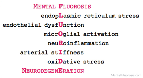
94 references are cited. 28 of the studies
were published in 2017 & 2018.
(See 2019 & 2020 research.)
Fluoride and ER Stress Dental Fluorosis: Researchers at the Forsyth Institute in Massachusetts, a fluoride research center for the past century, found that fluoride initiates an ER stress response in ameloblasts that interferes with protein synthesis and secretion culminating in dental fluorosis. Beginning with the lowest dose tested, they observed an increase in the magnitude of ER stress with increasing doses of fluoride. [Sharma R, et al. 2008] Ameloblasts are cells present during tooth development that secrete large amounts of proteins that later mineralize to form tooth enamel. Skeletal Fluorosis: Fluoride causes ER stress during osteoblast maturation. [Zhou et al. 2013] Osteoblasts are cells that secrete the protein matrix for bone formation. The severity of osteofluorosis is associated with accumulation of fluoride in bone, in a dose-dependent manner. [Liu L, et al. 2015] Pineal Fluorosis: Because it is not protected by the blood-brain barrier, the pineal gland is exposed to fluoride in the bloodstream. Its fluoride concentrations are positively related to calcium concentrations. By old age, the fluoride/calcium ratio in the human pineal gland is higher than in bone. [National Research Council (1) 2006; Luke 2001] (See MacArthur 2013) Animal research has found fluoride concentrations four times higher in the pineal gland than in the skull or the brain. [Kalisinska et al. 2014] Soft Tissue Fluorosis: Fluoride exposure induced excessive ER stress and associated apoptosis (cell death) in the hippocampus of rats, resulting in histological and ultrastructural abnormalities that impaired learning and memory capabilities. [Niu et al. 2018] Fluoride has also been shown to induce ER stress that causes cell death in spleen lymphocytes [Deng et al. 2016] and in immature sperm cells. [Yang Y, et al. 2015] Placental Fluorosis: Women who drank water fluoridated at 1.0-1.2 ppm (parts per million) averaged 2 ppm fluoride in their placentas nearly three times higher than in women who drank unfluoridated water. [Gardner et al. 1952] (See MacArthur 2015) |
ER Stress and Neurodegeneration The endoplasmic reticulum is the main compartment involved in protein folding and secretion and is drastically affected in Alzheimer's disease neurons. [Gerakis et al. 2018] Accumulating evidence indicates that chronic ER stress is of paramount significance in development and progression of many neurodegenerative diseases whose pathology includes accumulation of misfolded proteins in the brain. [Mahdi et al. 2016] In stroke and Lou Gehrig's disease, chronic activation of ER stress is considered as main pathogeny. [Zhang et al. 2015] ER stress and accumulation of several types of misfolded proteins (β-amyloid, Tau, alpha-synuclein, etc.) are associated with most of the brain pathology processes observed in the development of Alzheimer's disease. [Li JQ, et al. 2015]
ER stress also mediates the pathogenesis of psychiatric diseases, such as depression, schizophrenia, sleep fragmentation, and post-traumatic stress disorder. Inhibiting specific causes of ER stress may help prevent neurodegeneration. [Xiang et al. 2017]
Note: ER stress is a central mechanism in the pathogenesis of preeclampsia, the life-threatening pregnancy complication caused by the abnormal placenta. [Burton et al. 2011] |
ER Stress References Cited
Fluoride and Endothelial Dysfunction Sodium fluoride causes dramatic endothelial cell barrier dysfunction. [Wang P, et al. 2001] Sevoflurane, a Fluorinated Anesthetic
Sevoflurane is associated with high rates of post-operative delirium, a common and often fatal disorder affecting as many as 50% of older people during surgery or hospitalization. The severity of post-operative delirium correlates directly to the severity of later cognitive impairment and decline. [Institute for Aging Research 2017] Inhibiting ER stress during sevoflurane anesthesia will be a potential method to prevent later learning and memory deficits in adulthood. [Zhu G, et al. 2017] Another drug that increases blood fluoride levels is 5-fluorouracil, which also causes endothelial cell dysfunction. [Hull et al. 1988; Altieri et al. 2017] Fluoroquinolone antibiotics induce cytotoxicity to endothelial cells, depending on drug concentration and duration of exposure. [Bezwada et al. 2008] Fluoride and/or arsenic induced significant increase in endothelial cell death and inflammation, mechanisms of vascular toxicity. [Ma et al. 2017] Note: Amniotic tissue cultures treated with sodium fluoride have significantly higher expression levels of soluble Endoglin. [Tskitishvili et al. 2010] |
Endothelial Dysfunction and Neurodegeneration Microvascular endothelial cells play a critical role in brain development, maturation, and homeostasis. Acute or chronic insults cause brain endothelial cell dysfunction and damage. Loss of BBB integrity is a common pathological finding in many psychiatric disorders including autism and schizophrenia. BBB dysfunction leads to infiltration of peripheral material, culminating in neuroinflammation and oxidative stress. [Kealy et al. 2018] Endothelial cell dysfunction in the blood-brain barrier is the first pathological change in the development of small vessel disease, a major cause of vascular dementia in older people that triples the risk of stroke and contributes to Alzheimer's disease. [Rajani et al. 2018; Kelleher et al. 2013] A common mechanism in age-related diseases like dementia, cancer, and diabetes is the accumulation of dysfunctional endothelial cells that are not just an effect of aging. It's a reason why we age. [University of Exeter 2018] ER stress induces endothelial cell dysfunction in the brain and blood vessels. [Rizzo et al. 2010; Soejima et al. 2018] Chronic activation of ER stress in endothelial cells leads to increased oxidative stress and inflammation and often results in cell death. [Lenna et al. 2014] Note: In the complex pathogenesis of preeclampsia, endothelial dysfunction remains the most agreed-upon central mechanism involved. [Meeme et al. 2017] A marker of endothelial dysfunction is soluble Endoglin, whose levels are elevated in preeclampsia. [Al-Jameil 2014] |
See also the page about endothelial cell dysfunction.
Endothelial Dysfunction References Cited
Fluoride Activates Microglia and Neuroinflammation Fluoride can cause fluorosis and irreversible brain damage. The neuroinflammation process is meditated by microglia: activated microglia release pro-inflammatory cytokines and neurotoxic molecules. [Chen et al. 2017] Fluorine affects oxidative stress, glial activation and inflammation in the central nervous system, which leads to neurodegeneration. [Dec et al. 2017] Fluoride induced neuron apoptosis and inflammatory factors by activating microglia in rat brain. [Yan N, et al. 2016] When rats were treated for 30 days with fluoride in their drinking water, it caused an increase in microglial activation and neuroinflammation. [Akinrinade et al. 2015] In the central nervous system, inflammatory cytokines are released from activated microglial cells induced by fluoride. [Yan L, et al. 2013] Fluoride exposure activated microglia and promoted neuroinflammation in the hippocampus of rats sub-chronically exposed to fluoride in their drinking water, which affected synaptic plasticity by decreasing levels of PSD-95. [Yang L, et al. 2018] In rats that drank water with added fluoride for several months, the fluidity of brain synaptic membranes and the expression level of PSD-95 decreased in a dose-dependent manner. [Qian et al. 2013; Zhu W, et al. 2011] Rats anesthetized for 4 hours with 2.5% Sevoflurane a general anesthetic that significantly increases blood fluoride levels showed longterm deficits in hippocampal function and decreased hippocampal PSD-95 expression. Seven weeks after exposure, they had significant spatial learning and memory impairment. [Wang SQ, et al. 2013] (Sevoflurane studies cited above in Fluoride and ER Stress.) |
Microglial Activation, Neuroinflammation and Neurodegeneration Growing evidence links microglial activation to the development and progression of a variety of neurodegenerative disorders, including Alzheimer's, Parkinson's, Huntington's, and Lou Gehrig's diseases. A common hallmark in the pathogenesis is neuroinflammation. [Hickman et al. 2018] "Microglia are the linchpin of the neurodegenerative process" an appealing target of efforts to prevent cognitive decline in neurodegenerative diseases. [Washington Univ. School of Medicine 2019] Inflammatory cues trigger microglial cells to release the protein ASC. Specks of ASC promote aggregation and deposition of β-amyloid protein, a hallmark of Alzheimer's disease. [Ranshoff 2017] This review looked at the role of ER stress signaling in brain-associated immune cells and the implications to neuroinflammation and development of neurodegenerative diseases. [Garcia-Gonzalez et al. 2018] Chronic neuroinflammation is sustained by persistent and aberrant microglial activation in the brain. [McKenzie et al. 2017]
|
Microglia and Neuroinflammation References Cited
Fluoride and Arterial Stiffness Fluoride accelerates arterial calcification and stiffening that lead to hypertension. In people with fluorosis, the elastic properties of the ascending aorta are impaired. [Varol et al. 2010; Machoy-Mokrzynska 2004] Hypertension prevalence increased 22% when fluoride levels in drinking water increased from 0.84 to 1.55 mg/l. [Sun et al. 2013] PET/CT scans show a significant correlation between fluoride uptake and calcification in most of arterial walls. The coronary fluoride uptake value in patients with cardiovascular events was significantly higher. [Li Y, et al. 2012] Fluoride is readily incorporated into the crystalline structure of bone and will accumulate over time, resulting in arthritic symptoms of "joint stiffness and pain." [National Research Council (2) 2006]
Data from nearly 8,000 medical practices in England showed that hypothyroidism was positively associated with fluoride levels in patients' drinking water. Those in a fluoridated area had twice the hypothyroidism prevalence of a nonfluoridated area. [BMJ 2015] A Canadian study published in December 2018 found that the risk of hypothyroidism increases in adults who have iodine deficiencies and higher urinary fluoride levels. [Malin et al. 2018] Another 2018 Canadian study found that urinary fluoride levels are almost two times higher for pregnant women living in fluoridated regions compared with nonfluoridated regions. [Till et al. 2018] Racial Susceptibility to Fluorosis
|
Arterial Stiffness and Neurodegeneration Arterial stiffness correlates with the pathogenesis of a large spectrum of vascular disorders including hypertension, kidney dysfunction, cerebral small vessel disease, and decreased cognitive function, such as Alzheimer's disease. Worldwide, it's estimated that 9.4 million deaths per year are related to arterial hypertension. [Mikael et al. 2017; Singer et al. 2014] Hypertension causes stiffening and loss of elasticity in blood vessels, which affects the movement of interstitial fluid out of brain cells. This hinders clearance of the Alzheimer's pathology protein β-amyloid from the brain. [McMurray 2019] Aortic stiffness reduces the buffering of pulsatile blood flow, exposing cerebral small arteries to microvascular damage. [Kennedy et al. 2018] Arterial stiffness is determined by pulse wave velocity (PWV), a noninvasive measure of the speed at which the blood pressure pulse travels through the arteries. Older adults with high PWV readings were 60% more likely to develop dementia. Even after adjusting for age, sex, race, education, BMI, and anti-hypertensive medication, arterial stiffness was significantly associated with increased risk of dementia. [Cui et al. 2018] Aortic stiffening is an early trigger of abdominal aortic aneurysm. [Raaz et al. 2015] Impaired elasticity of the ascending aorta is associated with subclinical hypothyroidism. Higher TSH levels, a measure of hypothyroidism, are positively correlated with aortic stiffness. [Yurtdas et al. 2013] Subclinical hypothyroidism in pregnant women is associated with decreased IQ of their offspring. The decline in iodine intake in the US could contribute to increased toxicity of fluoride for some individuals. [National Research Council (3) 2006] Racial Susceptibility to Cardiovascular Disease
Stiffness of the placenta is significantly higher in preeclampsia, which is characterized by increased arterial stiffness. [Kilic et al. 2015; Kaihura et al. 2009] Patients with mild preeclampsia had impaired aortic elasticity. [Ciftci et al, 2014] |
Arterial Stiffness References Cited
Fluoride and Oxidative Stress It's long been recognized that fluoride increases ROS and causes oxidative stress. A PubMed Title/Abstract search for fluoride and oxidative stress lists over 300 studies published since 1990. For example: "Over uptake of fluoride for a long term could cause potential increase in the level of oxidative stress in the brain tissue." [Shao et al. 2000] "Fluoride at a dose encountered in drinking water in contaminated areas exerts an adverse effect on the male reproductive system and this effect is associated with indicators of oxidative stress." [Ghosh et al. 2002] Flash Forward A major review described the impact of fluoride-induced oxidative stress and inflammation in the pathogenesis of Alzheimer's disease, as well as a mechanism for its initiation by fluoride. [Goschorska et al. 2018 Dec 9] Fluoride Decreases Glutathione Levels
The neurotoxicity of fluoride is associated with oxidative stress due to imbalance between production and removal of ROS, including reduction of total glutathione content. [Wu J, et al. 2015] Increased levels of fluoride in drinking water are associated with reduced levels of glutathione and with an increased prevalence of carotid artery atherosclerosis in adults. [Liu H, et al. 2014] Glutathione content decreased markedly in fluoride-treated groups. [Shuhua et al. 2012] Fluoride Decreases Acetylcholine Levels
A study published in December 2018 explores how fluoride reduces acetylcholine levels in the brain by suppressing antioxidant activity. [Goschorska et al. 2018 Dec 20] |
Oxidative Stress and Neurodegeneration It's also well known that oxidative stress is involved in neurodegeneration. A PubMed Title/Abstract search for oxidative stress and neurodegeneration lists over 3,900 studies published since 1990. For example: "Because of the scavenging activity of glutathione against accumulation of oxygen radicals, its decrease in the brains of parkinsonian patients has been interpreted as a sign of oxidative stress." [DiMonte et al. 1992] "Oxidative stress and excessive activation of glutamate receptors... provide a final common pathway for cell vulnerability in the brain." [Coyle et al. 1993]
Glutathione Depleted in Alzheimer's
"If routine non-invasive tests for lower levels of glutathione in the hippocampus regions are performed, we might be able to mitigate the advancement of Alzheimer's disease by providing glutathione supplements." [Mandal et al. 2018] In a March 2019 editorial, Dr. Mandal highlighted the need for glutathione supplementation in mild cognitive impairment and Alzheimer's. [Mandal et al. 2019] Maintaining glutathione levels may provide a therapeutic treatment for Parkinson's disease. [Smeyne & Smeyne 2013] Acetylcholine Decreased in Alzheimer's
|
Oxidative Stress References Cited
October 2019:
Published in the Townsend Letter: The Examiner of Alternative Medicine